
High Resolution AFM with Bruker PeakForce Tapping
PeakForce Tapping
PeakForce Tapping is a powerful AFM (Atomic Force Microscopy) technology, unique to Bruker microscopes. It enables extremely high resolution imaging and extends AFM applications into areas that were not previously possible. It also enables nanoscale properties to be mapped simultaneously alongside imaging.
PeakForce Tapping is a unique feature of Bruker AFM microscopes. Blue Scientific is the exclusive distributor of Bruker AFM in the Nordic region (Sweden, Norway, Denmark and Finland). If you have any questions or if you would like a quote, please contact us:
Contact us on +44 (0)1223 422 269 or info@blue-scientific.com
View Bruker Atomic Force Microscopes
Highest Resolution Imaging
With PeakForce Tapping, probe-to-sample interaction is controlled precisely, for the lowest possible imaging force. This extra level of control results in more consistent and higher resolution AFM imaging, with piconewton (pN) force sensitivity. Using lower forces has the additional benefit of preserving the probe shape for longer life and more consistent imaging. The technique is suitable for a wide range of sample types, from soft biological samples to hard materials.
Quantitative Nanoscale Mapping
Using Bruker atomic force microscopes, nanoscale imaging can be combined with quantitative electrical, mechanical, biological and chemical property mapping, providing you with a whole new range of data. More details about specific modes is below.
Easy to Use
PeakForce Tapping is easy to use, and works with Bruker’s ScanAsyst image optimisation software. ScanAsyst automates the process, so that users of all experience levels can acquire high quality images. The software is self-optimising, with algorithms that continuously monitor image quality and adjust parameters accordingly. Simply select the scan area and size for your sample, in air or fluid.
Which AFMs Feature PeakForce Tapping?
- Dimension FastScan
- Dimension FastScan Bio
- Dimension Icon
- Dimension Icon-Raman
- Inspire
- BioScope Resolve
- MultiMode 8
- Dimension Edge
How Does it Work?
The probe periodically taps the sample. The pN-level interaction force is measured by the deflection of the cantilever. With superior force control, the feedback loop keeps the peak force constant, down to 10 pN. This is significantly lower than other modes, which are typically around ~1 nN. The technique works in both air and fluid.
PeakForce Tapping AFM Modes and Applications
PeakForce Tapping technology enables and enhances other correlative and quantitative mapping techniques. This opens up a whole range of new possibilities in topographical, mechanical, biological, electrical and chemical applications at the nanoscale.
PeakForce QNM – Quantitative Nanoscale Mechanical Characterisation
Ideal for materials science – simultaneously map and distinguish nanomechanical properties while imaging the topography of your sample, all at atomic scale resolution. Study the variation and position of mechanical properties across a surface, with no ambiguity about the source of image contrast, including modulus, adhesion, dissipation and deformation.
In life science research, correlate sub-molecular AFM with quantitative mapping of mechanical, chemical and biological interactions. Simultaneously image and map the mechanical properties of live cells, viruses and tissues.
Left: Modulus image showing the transition between the ULDPE tie layer to the PS/LDPE sealant layer of a cross-sectioned packaging material. Image size 3 um.
PeakForce TUNA – Current Mapping on Fragile Samples
Study the conductivity of fragile samples such as organic photovoltaics, conductive nanotubes and nanoparticles. PeakForce TUNA does not suffer from the the limitations of conventional contact-mode-based conductive AFM, which can cause sample damage or probe tip contamination from CAFM lateral forces.
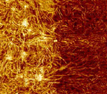
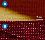
Left: Height (A) and current (B) maps of a carpet of vertical carbon nanotubes, which are impossible to image with contact mode. Image size 1 μm.
PeakForce Kelvin Probe Force Microscopy – Artefact-Free Potential Contrast
Measure surface potential with greater spacial resolution and accuracy than traditional KPFM (Kelvin Probe Force Microscopy).
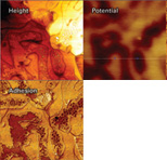
Left: Height, adhesion, and surface potential images of Sn-Pb obtained with PeakForce KPFM. Image size 4 μm.
PeakForce IR – Quantitative Nanochemical Characterisation
Acquire more information than traditional AFM, sSNOM and photothermal techniques, from a wider range of sample types. Acquire instantly correlated chemical and nanomechanical information, sich as modulus, adhesion and deformation. Identify material types and detect variations in film thickness with molecular monolayer sensitivity. Characterise powders, polymer brushes, rubbery components, metals, semiconductors, ceramics, graphene and 2D materials.
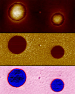
Left: Chemical and nanomechanical information for a polystyrene/LDPE blend. Image size 1 μm x 2 μm.
PeakForce Capture – Nanomechanical Data for Every Pixel
PeakForce Capture records force curves for every pixel of your image, as well as calculated property channels. Force curves from the PeakForce QNM image are saved alongside the image file in a proprietary 3D data cube format. Capture unexpected events with high sensitivity, which may not be recorded by other techniques.
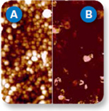
Left: Height (A) and stiffness (B) maps of calcite, revealing increased contact stiffness for alternate rows of atoms.
ScanAsyst – Self-Optimising AFM
ScanAsyst makes it easier to acquire high quality images, by using intelligent algorithms to continuously adjust settings and parameters for the best possible quality. Image live cells and molecules, with high resolution cellular detail.
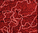
Left: DNA imaged using ScanAsyst on a MultiMode 8. Image size 1 μm.
Contact us on +44 (0)1223 422 269 or info@blue-scientific.com
View Bruker Atomic Force Microscopes