
Microplastics Analysis with Raman Spectroscopy
Raman spectroscopy is ideal for studying microplastics and analysing bulk plastic contamination. Raman is a non-destructive optical scattering technique that can identify particles and fragments quickly, with automated features for reliable results:
- Locate fragments
- Identify polymers present
- Measure particle size and composition
Blue Scientific is the official distributor for Renishaw Raman in the Nordic region (Norway, Sweden, Denmark, Finland, Iceland). For more information or quotes, please get in touch.
Renishaw InVia
More posts about Raman
Contact us on +44 (0)1223 422 269 or info@blue-scientific.com
Benefits of Raman Spectroscopy
- Analyse a range of sizes of particles, from 1 µm up to several millimetres across
- Automated measurements for consistent data
- Wide range of analytical tools:
- Identify particles with spectral databases
- Measure composition, size and shape
- Analyse particles on a wide range of substrates with advanced focus-tracking
Renishaw InVia
The Renishaw InVia is a research-grade confocal Raman microscope:
- Acquire detailed chemical images and highly specific data from discrete points.
- Analyse both large volumes and minute traces of materials.
- Easy-to-use software for data collection, analysis and display.
Automated Workflow
With the InVia’s software, you can build automated workflows in the software for fast analysis. A typical workflow consists of:
- Deposition – Deposit the microparticles on a substrate.
- Loading – Place the substrate on the microscope stage.
- Optical imaging – A high resolution image is built from multiple high-magnification images. Where the optical contrast is limited, plastic regions can be determined by laser mapping.
- Particle determination – The image is analysed automatically to locate the plastic particles.
- Raman analysis – Each particle is then analysed, either by:
- A single spectrum from the centre of the particle
- Mapping the entire particle eg to check for a mixture of polymer types
- LiveTrack focus-tracking can be used for rough or uneven surfaces
- Analysis – The spectra are matched to a database of polymers. You can also customise the database by adding to it, or create your own.
Example: Analysing Microplastics in Sea Water
In this example, microplastic particles from sea water were deposited on a mirror-polished stainless steel slide. This substrate provides high optical contrast and does not affect the polymer spectrum.
While this is a flat substrate, it’s also possible to analyse rough and uneven surfaces with Renishaw’s LiveTrack focus-tracking technology.
Optical Imaging
Below is the high resolution optical microscope image of the substrate (1.8 mm × 1.1 mm). Contamination usually appears as large particles, so these areas were selected for further analysis. It’s also possible to select smaller particles for more in-depth studies.
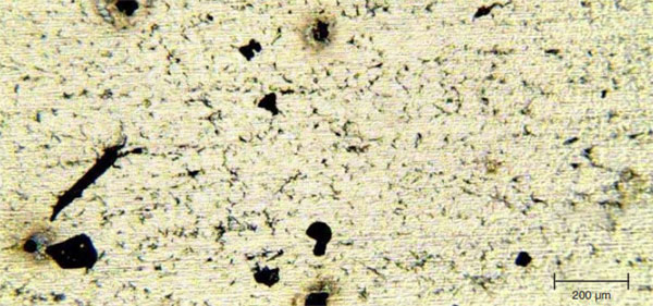
Optical microscope image of microplastics on mirror-polished stainless steel substrate.
Particle Determination
Particles are located automatically by the software, shown in the image below. Five have been assigned numbers for further investigation.

Particles are located automatically by the software for further analysis.
Identification
Each particle was mapped and compared to a database of polymers and inorganic materials for identification:
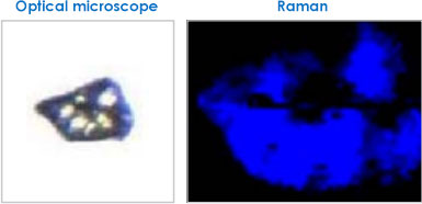
Particle 1: Mostly polypropylene (blue in the Raman image)
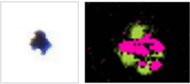
Particle 2: Hematite (pink) and another iron-based inorganic (lime green)
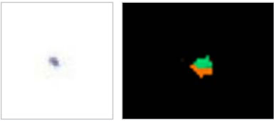
Particle 3: Anatase (green) and quartz (orange)
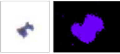
Particle 4: Silica (purple)
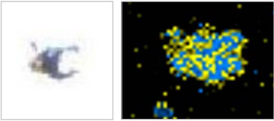
Particle 5: Mixture of PTFE (blue) and an unidentified
fluorescent material (yellow), probably a dye.
Two out of the fourteen particles were found to be microplastics (polypropylene and PTFE). The others were identified as inorganic and mineral-based species, including hematite, pyrrhotite, anatase, quartz and silica.
Further Information
Blue Scientific is the official Nordic distributor of the Renishaw InVia Raman microscope. If you have any questions or if you’d like a quote, please get in touch:
Contact us on +44 (0)1223 422 269 or info@blue-scientific.com
Renishaw InVia
More posts about Raman