16 Mp CMOS camera
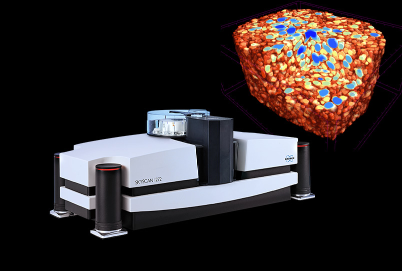
The Bruker SkyScan 1272 is a desktop micro-CT scanner (3D X-ray microscope). A state-of-art 16 megapixel CMOS X-ray detector delivers superior resolution. With a native resolution of up to 11200 x 11200 pixels per slice, you can zoom in to any part of the 3D volume without re-scanning. The compact system can be placed on any lab desk, requiring only a standard power supply.
The variable detector position can move closer to the sample, increasing intensity and reducing scan times.
For unattended, high-throughput analysis.
Optimise settings with a single click for the best results.
If you’re new to micro-CT or short on time, use Genius Mode to select the best parameters for your sample. Magnification, energy, filters and exposure time are automatically selected and optimised. These can also be adjusted manually to get exactly the results you want.
Both the sample and large-format CMOS camera can be positioned close to the X-ray source, with variable detector geometry. This significantly increases the intensity, making the SkyScan 1272 up to 5 times faster compared to conventional scanners with a fixed camera position.
The desktop system can be placed on any lab desk and has a footprint of only 0.6 m² (116cm x 52cm). It required only a standard domestic power socket – no water chiller or additional compressor. The industry-grade sealed X-ray source is maintenance-free, saving maintenance costs over time.
An optional sample changer is available for up to 16 samples, so the SkyScan 1272 can run unattended, increasing throughput for QC and routine analysis. The sample changer can handle samples up to 96mm in diameter, and any mixture of sizes.
With external access, samples can be can be replaced at any time, without interrupting scanning. New samples are detected automatically, and LEDs indicate the status of every scan.
In-situ testing stages are available, which communicate with the stage automatically, with no cable connections.
We offer a range of instruments for elemental and mineral analysis, mapping and imaging in geology and geoscience.
Systems for food process and quality control and analysis.
How Bruker's Genius-Mode automatically optimises micro-CT imaging parameters for the best quality results.
A new method for automatic trabecular-cortical separation in micro-CT called the morphological escalator.
How to analyse individual and multiple fibres in natural and synthetic materials using micro-CT imaging.
How multi-scale micro-CT analysis can be used to achieve the best micro-CT image quality, using geological samples as an example. How to get the best results from your micro-CT scanner, and achieve the highest resolution. If you have any questions about how to best use micro-CT analysis for your research, please get in touch: Contact us on 01223 […]
Instrumentation and techniques for studying foam with micro-CT. Analyse the internal structure, map morphometric properties & perform dynamic in-situ experiments (tensile, compression & modified temperature).
An overview of Bruker SkyScan micro-CT applications in life science, including in vivo/ex vivo, dental and bone research.
X-ray micro-computed tomography (μCT) is a non-destructive technique for studying internal micro-structure in 3D – with sub-micron resolution. It’s also known as X-Ray Microscopy (XRM). In the food industry, you can investigate the relationship between structure and properties in product development, as well as gaining insights into shelf life, etc. Analyse samples of any shape, […]