Micro-CT (also known as XRM or X-Ray Microscopy) is a non-destructive 3D imaging and analysis technique, for studying the internal structure of all types of samples, with diameters from 1 mm to 20cm and full drill cores. Take virtual slices through objects and build stacks for 3D volume rendering.
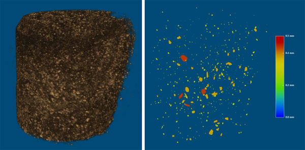
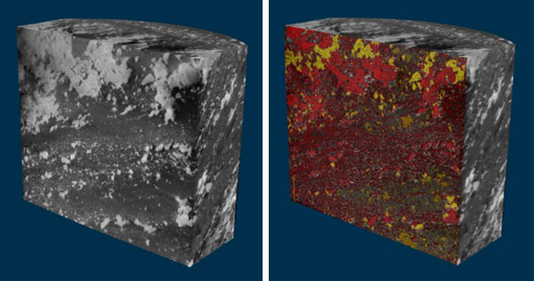
Left: 3D mineral distribution of gold deposits.
See inside your sample with non-destructive 3D X-ray imaging
Applications
Micro-CT is used in all areas of geology including:
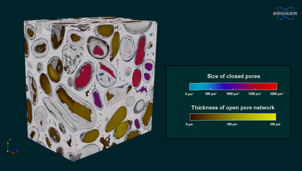
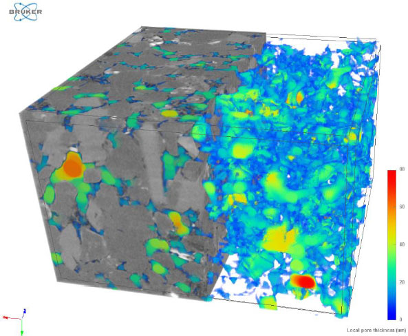
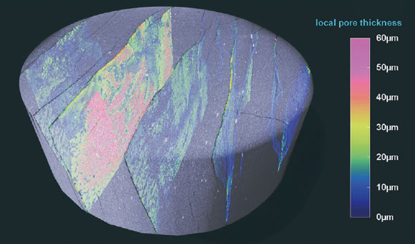
- Porosity
- Micro-structure
- Composition
- 3D mineral distribution
- 3D analysis of shape and morphometries, including grains, sedimentary patterns, fossils, etc
- Multi-scale analysis
- Dynamic processes eg weathering, movement of fluids, crystallisation
All fields of geoscience:
- Oil and gas
- Sedimentology
- Structural geology
- Construction materials
- Geochemistry
- Paleontology
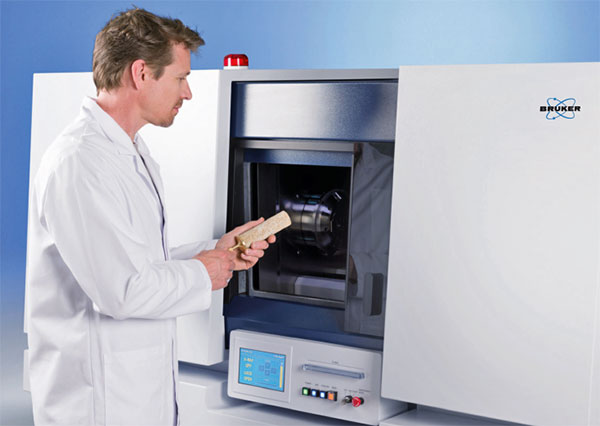
Scanning a sample with the Bruker SkyScan 2214
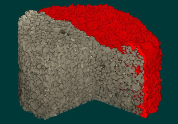
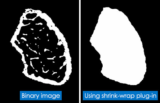
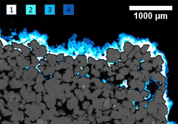
Select Complex Regions of Interest
ROIs are rarely regular, especially in rock samples. Bruker’s micro-CT software has powerful features for making selections and extracting the data you need.
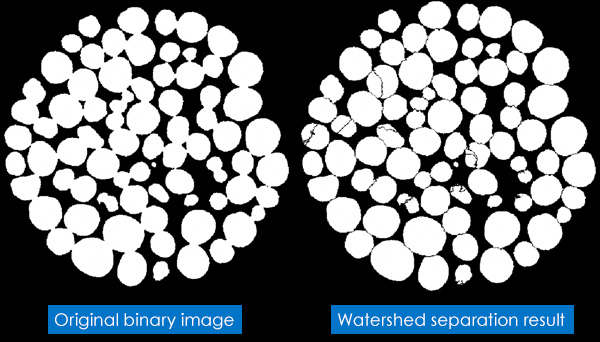
Measure Grains Individually
With Bruker’s watershed separation algorithm, you can analyse grains and particles individually, even if they’re closely packed.
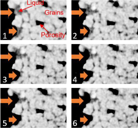
Observe Dynamic Processes
Image dynamic processes with step-by-step timelapse and in real time methods. This is known as 4D micro-CT.
Multiscale Analysis
Extract and combine information from various length scales to quantify and model the overall sample behaviour.
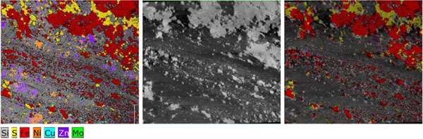
Complementary Technologies
The data acquired from X-Ray Microscopy adds value to other analytical techniques, including micro-XRF, XRD and optical / electron microscopy. Data can be overlaid to create 3D mineral maps and more.
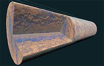
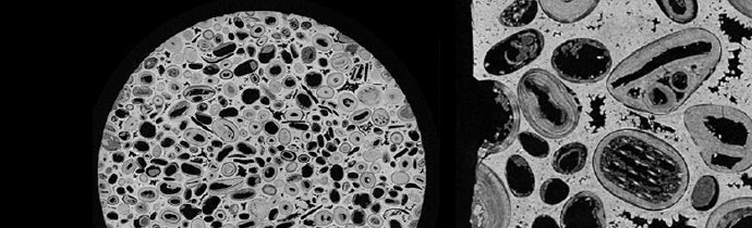
4mm Chalk Sample
Instruments
Bruker SkyScan 1275
Automated, benchtop micro-CT with self-optimising features, so you can scan at the press of a button. Extremely fast image reconstruction – a practical way to add micro-CT capabilities to your facility.
Bruker SkyScan 2214
Multiscale nano-/micro-CT covering the widest range of resolutions. Sub-micron resolution and the world’s fastest hierarchical 3D reconstruction.
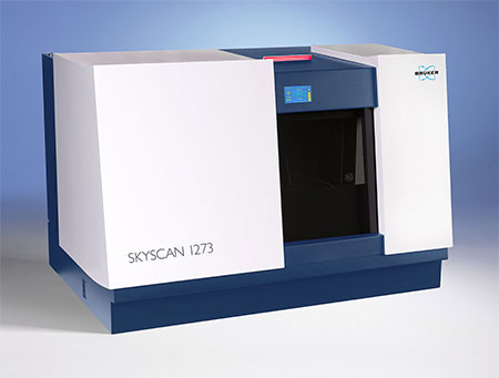
Bruker SkyScan 1273
Benchtop XRM scanner for large and/or heavy objects up to 20kg. Ideal for rock samples, drill cores, etc.