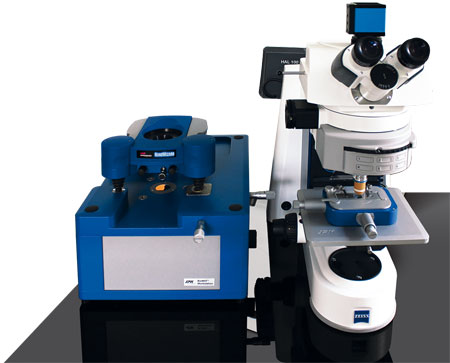
BioMAT Workstation
Upright optical microscopy and AFM for opaque samples
The Bruker JPK BioMAT Workstation solves one of the biggest problems in correlative microscopy of non-transparent samples: providing simultaneous access for both techniques. The system enables both upright optical microscopy and AFM to operate at the same location, with no compromise to either.
- Completely integrated system
- High resolution imaging and characterisation of the nano-mechanical and nano-electrical sample properties
- Combine AFM with virtually all upright microscopy techniques, including brightfield, DIC, fluorescence, confocal laser scanning, FLIM FRET, FRAP, FCS, IR and Raman
- Suitable for operation in liquids
- Flexible concept with shuttle stage
Contact us for more information and quotes:
+44 (0)1223 422 269 or info@blue-scientific.com
BioMAT Shuttle Stage
The key component of the BioMAT Workstation is a portable shuttle stage that carries the sample. This transfers the sample between the optics’ field of view and the AFM scan range without losing its position. This transfer can be repeated as many times as you need to, for the sequential measurement of optics and AFM in time lapse studies.
This is even compatible with in-fluid operation, using dipping lenses for the upright microscope.
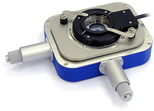
BioMAT shuttle stage with the petri dish heater
Applications
With the JPK BioMAT Workstation you can study samples on non-transparent substrates, such as tissue sections and bacterial growth on metallic surfaces.
In addition to life science applications, the combination of AFM with optics enchances surface chemistry and polymer research, as well as nano-electrical measurements on semiconductors and micro-mechanical systems (MEMS/NEMS).
- Biochips eg DNA and protein chips
- Cell-electronics interfaces, cell chips and patterned substrates for cell growth
- Tissue engineering and implants
- Bacterial or yeast studies on non-transparent substrates like metals or plastics biofouling
- Bionics studies
- Plant biology
- Nanostructured surfaces from PDMS or other imprints
- Micromechanical (MEMS/NEMS) systems
- Nanoparticles, powders, foams, paintings and thin films
- Organic coatings on metals, plastics or silicone substrates for biocompatible surfaces
- Nano-electrical measurements of electrical devices
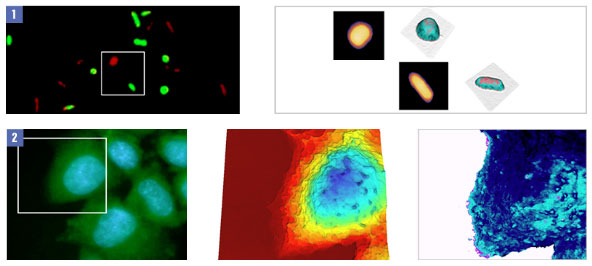
1: Gram negative Paracoccus Seriniphilus bacteria. 2: Live CHO cells on gold electrode, measured using the BioMAT PetriDishHeater
Combine with Confocal Laser Scanning Microscopy
By combining high resolution AFM with the latest optical microscopes you can build new experiments for quick and precise sample surveys. These can be combined with nanometre-scale, quick tests of geometry and secondary sample properties including mechanical, electrical and magnetic characteristics.
More details are available in this note.
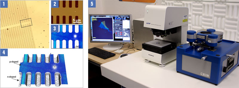
1: D profile of doped (p, n) semiconductor structure. 2: Corresponding AFM topography. 3: EFM image of the marked region. 4: 3D Overlay of AFM topography and EFM.