Combine Raman with other Microscopy Techniques (SEM, AFM, IR & more)
How to combine results from Raman spectroscopy with other microscopy techniques, including SEM, AFM, fluorescence, IR, optical microscopy and more. Gain greater insights into your sample by combining Raman images with Renishaw’s new Correlate software module.
Blue Scientific is the official distributor for Renishaw Raman in the Nordic region (Norway, Sweden, Denmark, Finland, Iceland). For more information or quotes, please get in touch.
Renishaw Raman range
More articles about Raman
Contact us on +44 (0)1223 422 269 or info@blue-scientific.com
Follow @blue_scientificCombine Microscopy Techniques
Renishaw’s new Correlate software module enables you to combine imaging techniques for new insights from your microscopic methods.
Microscopy techniques are typically used separately in the lab. However, the results are often complementary, so combining them can lead to a better interpretation of your sample. Overlaying images from multiple techniques can give you a deeper understanding, as well as making the results easier to interpret.
New Renishaw Correlate Software
Renishaw’s new Correlate software module combines images from Raman spectroscopy with other imaging techniques. You can compare Raman results with other commonly used microscopic methods including:
- SEM,
- Fluorescence
- AFM
- Infrared
- Optical microscopy
Compatibility
The Correlate module is part of Renishaw’s WiRE 5.3 software. It can be used with these systems from Renishaw:
- inVia confocal Raman microscope
- Virsa remote sampling Raman analyser
- RA802 Pharmaceutical Analyser
- RA816 Biological Analyser
For information about the other techniques it can be used with, please get in touch.
Useful Software Tools
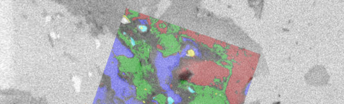
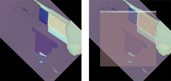
It’s easy to use; First record the coordinates of three or more reference points and data acquisition points on your sample. The Correlate module then guides you to these regions of interest, ensuring that data is acquired from the exact same locations. The images are then overlaid for interpretation.
- Coordinate Manager – Import and transform coordinates from commonly-used microscopy systems to the Raman system.
- Image Alignment Tools – Translate, rotate, adjust the size and aspect ratio of the overlay. You can also vary the transparency.
- Batch measurements – Automate the same Raman measurement at various positions on the sample.
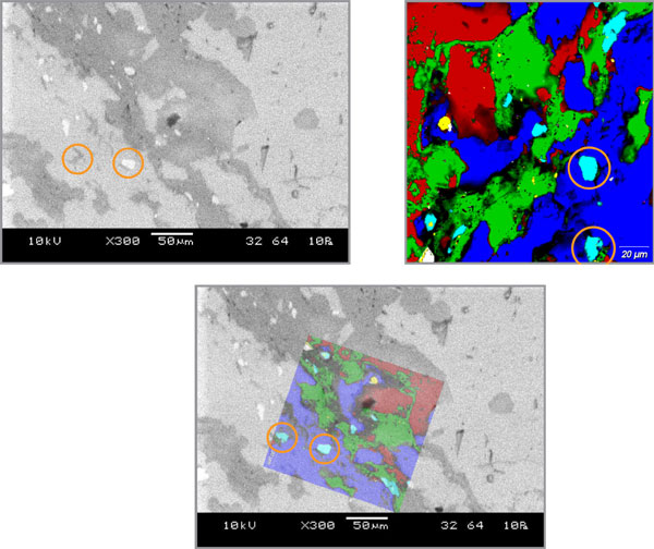
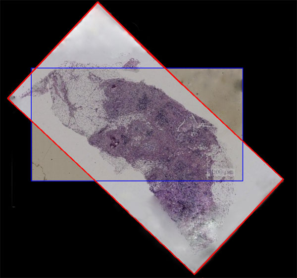
Example: Studying a Mineralogical Section
See how the new software can be used in this application note from Renishaw. Raman and SEM images of a mineralogical sample were combined to create more powerful data, incorporating both chemical and physical information.
More Information
Blue Scientific is the official distributor of Renishaw Raman in Scandinavia (Norway, Sweden, Denmark, Finland and Iceland). We’re available to provide quotes and answer all your questions – just get in touch:
Contact us on +44 (0)1223 422 269 or info@blue-scientific.com
Renishaw Raman