Renishaw Biological Analyser
Raman spectroscopy for biological and clinical research
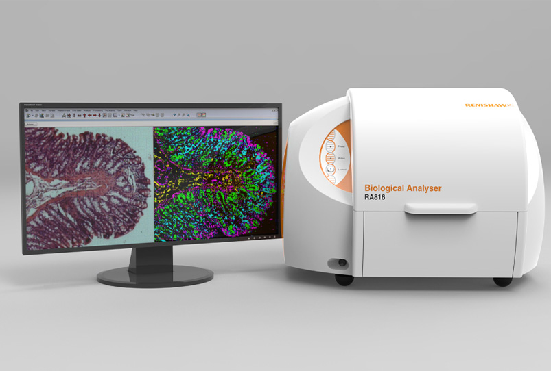
The Renishaw RA816 Biological Analyser is a Raman imaging system designed specifically for life science.
It delivers detailed information about the distribution and amount of biochemical species within biological samples, including tissue biopsies, tissue sections and biofluids.
- Non-invasive optical technique – maintaining your sample’s integrity
- Measure multiple molecular constituents in biological samples at once
- No sample staining or labelling
- High specificity – ideal for identifying early onset disease markers
- Raman images provide biomolecular information alongside spatial morphology
- Identify components in mixtures automatically with Renishaw’s new Spectrum Search module.
You can also combine results with images from other microscopy systems, includign AFM, SEM, IR, fluorescence and optical microscopes, using Renishaw’s Correlate software module – details here.
Contact us for more information and quotes:
+44 (0)1223 422 269 or info@blue-scientific.com
Designed for Clinics
The Renishaw Biological Analyser has been designed specifically to make Raman spectroscopy accessible in clinical research.
Translating Raman to the Clinic
- Build and validate pathology and disease models
- Validate models with new samples, to stratify and classify unknown tissues and biofluids
- Specially designed sample holders, accessories and substrates for bio-fluids/liquids and tissue sections
- Bulk tissue biopsy sample holder eliminates the need for tissue sample preparation (no cryosectioning or microtoming)
- Transfer disease models between instruments
Made for Demanding Applications
- Stable and repeatable results
- High throughput – high signal to noise spectral data and optimised substrates
- Maintain focus with LiveTrack™ technology
- Fast data collection with Streamline™ technology, without causing laser sample damage
- Low-cost, ultra-low background disposable mirror slides increase Raman signal and improve white light contrast
Easy to Use
- Easy to use software makes Raman spectroscopy accessible to all users, without compromising performance
- Experiment templates for various biological sample types
- Set up multiple measurements and leave to run unattended
Compact, Robust and Transportable
- Small footprint
- Move easily between lab and clinic
- Built-in performance checks to ensure accurate operation – including post-measurement checks
- No user alignment
Flexible Measurements
- Macro and high magnification modes, with digital zoom for various sample sizes
- Choose reflection or transmission illumination for the best image contrast
- Image tiling for large areas
Application Notes
These application notes and brochures are available from Renishaw’s website (registration or details required):
- RA816 Biological Analyser brochure
- Classifying brain glioma tumours
- Monitoring transdermal drug delivery in skin

Raman image of human bone cancer cells (osteosarcoma). Green = nuclei; Red = nucleoli; Cyan = membrane bound organelles; Yellow = cell body (thick region) Blue = cell body membranous area. Cell sample courtesy of Dr Frederick Coffman, Rutgers New Jersey Medical School, USA.

Human brain tissue. Left: white light. Right: Raman false-coloured composite score image of cerebellum whole follicle. Red = arbor vitae/white matter; Green = granule cell layer; Dark blue = molecular layer; Cyan = meninges (pia, arachnoid and dura mater).

Raman images of rat mandible, showing variation in surface topography. Renishaw’s LiveTrack technology maintains focus across the uneven surface.
Comments are closed.