Bone Morphometry in Micro-CT: Separating Trabecular and Cortical Bone
A new technique for automatic trabecular-cortical separation in micro-CT is now available, called the morphological escalator. This is useful in preclinical bone morphometry, and is available with Bruker SkyScan micro-CT scanners.
Blue Scientific is the official UK and Nordic distributor for Bruker Micro-CT. For more information or quotes, please get in touch.
Bruker Micro-CT systems
Contact us on +44 (0)1223 422 269 or info@blue-scientific.com
Follow @blue_scientificTrabecular Delineation in Micro-CT
Bone morphometry in rat and mouse pre-clinical imaging requires the medullary trabecular bone to be separated from the surrounding cortical bone at the endosteal boundary. One method of achieving this is by drawing regions of interest manually. However with advanced image processing, the trabecular delineation procedure can be automated.
One way of doing this uses morphological (erode, dilate, open, close), despeckle and bitwise (Boolean) image operations to separate trabecular from cortical bone at the rodent metaphyseal medulla. This technique is described in Bruker’s method note MN008: “Automated trabecular and cortical bone selection” (contact us for a copy).
Morphological Escalator
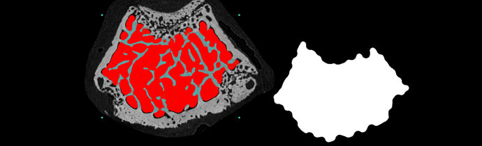
A new technique for trabecular delineation has been developed, called the morphological escalator. This automates the process, even in tricky regions where parts of the cortical wall are porous with only thin structures. This includes peripheral growth plate remnants downstream of the growth plate, often found in young, fast-growing rodents, such as the mouse femur metaphysis cross-section below.
Trabecular Bone Auto-Separation Methods
Trabecular bone auto-separation methods are based on these morphological operations:
- Erode (remove a pixel layer from the surface)
- Dilate (add a pixel layer to the surface)
- Open (erode then dilate)
- Close (dilate then erode)
Achieving a Clean Separation
A trabecular volume of interest (VOI) can be selected by binarising the medullary space and closing out the gaps from trabecular structures. However, there is often an overlap in thickness between the trabecular and cortical bone. This means that a single combination of opening and closing does not always achieve a clean separation of trabecular bone, often resulting in artefacts.
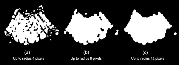
The morphological escalator solves this problem, using a series of open-close operations. It begins with a voxel radius of 1, then successively increases the voxel radius eg open 1 pixel; close 1 pixel; open 2 pixels; close 2 pixels, etc. The maximum value depends on the thickness of the trabecular structures.
The image below shows the results after opening and closing to a radius of 12 voxels. The example on the left used a single open-close morphological operation and on the right the stepwise morphological escalator.
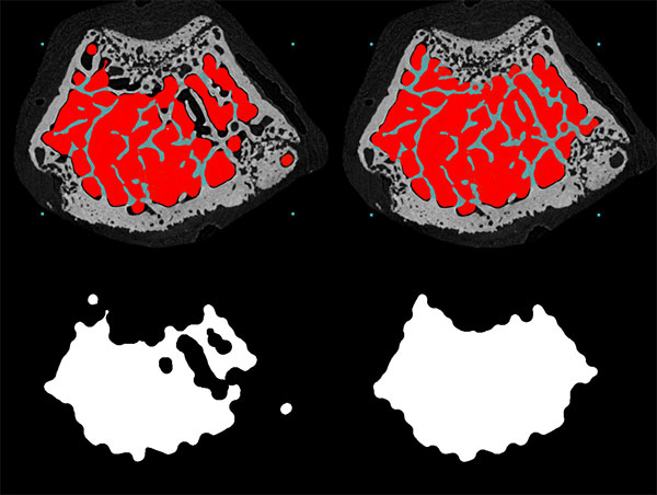
In the example above, the morphological escalator has successfully excluded peripheral remnants of growth plate from the trabecular VOI, including finely textured, porous regions of the cortical boundary. This restricts the analysis to mature secondary trabecular bone. This would not always be possible using standard trabecular-cortical separation algorithms.
How it Works
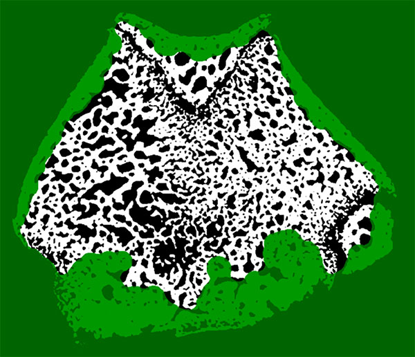
The image below shows how the morphological escalator works. Regions fill in as black or white progressively, according the local percentage volume and relative thickness of bone or soft tissue spaces.
When to use the Morphological Escalator
The morphological escalator works well for mouse bone with relatively sparse trabeculae, to exclude peripheral growth plate remnants.
For bones with highly dense medullary regions such as the example below, the percent trabecular volume can be above 50%. In such cases it’s more effective to use manual delineation.
Bruker Micro-CT Scanners
We offer the full range of microtomography systems from Bruker, including:
- SkyScan 1276
For in vivo small animals and biological samples - SkyScan 1275
Fast & self-optimising - SkyScan 2214
Multiscale nano-CT - SkyScan 1272
High resolution benchtop scanner - SkyScan 1273
High capacity for large samples - SkyScan 1278
Low dosage for in vivo applications
More Information
Blue Scientific is the official UK and Nordic distributor of Bruker Micro-CT. We’re available to answer all your questions – just get in touch: