
How micro-tomography can be used in biomedical research for sub-micron 3D imaging to study zebrafish morphology, bone densitometry, tissue, histology and more.

Using Bruker’s watershed algorithm to separate grain-based materials with X-ray micro-CT – ideal for powders, fibres, geological samples, pharmaceuticals and more.
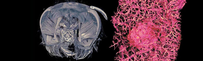
An overview of Bruker SkyScan micro-CT applications in life science, including in vivo/ex vivo, dental and bone research.
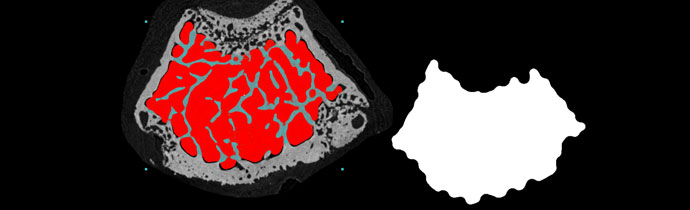
A new method for automatic trabecular-cortical separation in micro-CT called the morphological escalator.
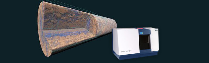
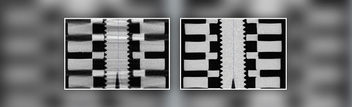
The Bruker SkyScan 1273 is a new micro-CT scanner for samples up to 500 mm length, 300 mm diameter and a maximum weight of 20 kg. This is a new standard for non-destructive testing (NDT) using a benchtop system. Blue Scientific is the official distributor for Bruker Micro-CT in the UK and Nordics (Norway, Sweden, […]
An explanation of helical micro-CT (spiral scanning) and how it’s used in materials science and orthopedic research.

In micro-CT 3D X-ray imaging, regions of interest are not always simple or regular. Often you need to select more complex regions. These ROIs can be selected automatically using two powerful plug-ins from Bruker: ROI shrink-wrap Primitive ROI Blue Scientific is the official distributor for Bruker Micro-CT in the UK and Nordic region. For more information or […]
Bruker are constantly updating their micro-CT software with new features and capabilities. Round-up of new tools and improvements in CTAn version 1.18.