The Bruker XRM User Meeting will be held online on 12th – 13th October 2021.
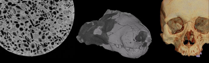
An overview of how microtomography is used in paleontology, to study the internal micro-structure of bones, teeth, fossils and more.

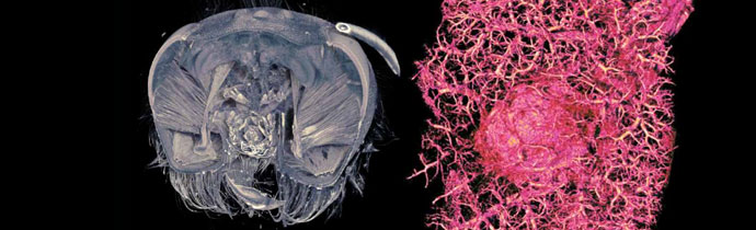
How micro-tomography can be used in biomedical research for sub-micron 3D imaging to study zebrafish morphology, bone densitometry, tissue, histology and more.

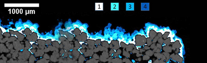
Using Bruker’s watershed algorithm to separate grain-based materials with X-ray micro-CT – ideal for powders, fibres, geological samples, pharmaceuticals and more.
An overview of Bruker SkyScan micro-CT applications in life science, including in vivo/ex vivo, dental and bone research.
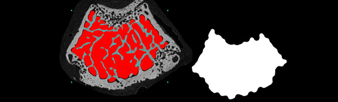
A new method for automatic trabecular-cortical separation in micro-CT called the morphological escalator.
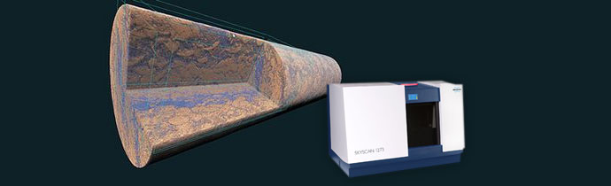
The Bruker SkyScan 1273 is a new micro-CT scanner for samples up to 500 mm length, 300 mm diameter and a maximum weight of 20 kg. This is a new standard for non-destructive testing (NDT) using a benchtop system. Blue Scientific is the official distributor for Bruker Micro-CT in the UK and Nordics (Norway, Sweden, […]
4D micro-CT (Micro-Computed Tomography or 3D X-ray imaging) involves imaging dynamic processes over time. This article explains step-by-step time-lapse and real-time methods, and how they can be used in geology and geoscience to image processes in rocks. These methods can also be used in other areas; If you have any questions just get in touch. Author: […]
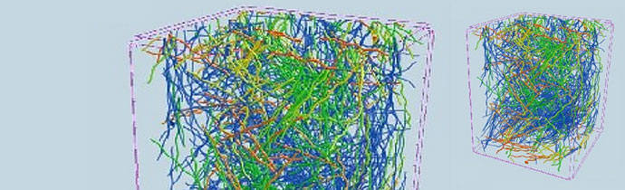
How to analyse individual and multiple fibres in natural and synthetic materials using micro-CT imaging.
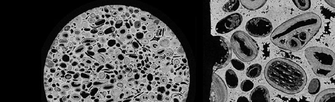
How multi-scale micro-CT analysis can be used to achieve the best micro-CT image quality, using geological samples as an example. How to get the best results from your micro-CT scanner, and achieve the highest resolution. If you have any questions about how to best use micro-CT analysis for your research, please get in touch: Contact us on 01223 […]